Bone Cross Section Labeled : Midsagittal Section Of Skull - A diagrammatic view of a cross section of bone.
byAdmin•
0
Bone Cross Section Labeled : Midsagittal Section Of Skull - A diagrammatic view of a cross section of bone.. Each system contains for a bone tissue engineering scaffold to be successful. The cross section of this circular cylinder is a circle. Red marrow fills the spaces in the spongy bone. (micrograph provided by the regents of university of michigan. Try pressing the section on the slide to ensure that the layer of glue is as thin as possible.
The previous image was correct, with one between the diaphysis and the head of the femur (which is an ossification center) and the other between the greater trochanter and the diaphysis. Cross section of bone labeled. The wider section at each end of the bone is called the epiphysis (plural = epiphyses), which is filled with spongy bone. It seems confusing and misleading. The wider section at each end of the bone is called the epiphysis (plural = epiphyses), which is filled internally with spongy bone, another type of osseous tissue.
Tooth Anatomy Medical Labeled Cross Section Chart With Enamel Dentin Pulp Gingiva Blood Vessels And Nerves Isolated Vector Illustration On White Background Stock Vector Adobe Stock from as2.ftcdn.net • learn about the materials that make up bone • label a cross section of bone. The image can be changed using any combination of the following commands. Each epiphysis meets the diaphysis at the metaphysis. See labeled cross sections of the human body now at kenhub. There are 7 main areas covered in the upper limb; Red bone marrow fills the spaces between the spongy bone in some long bones. The outside of a bone is covered in a thin layer of dense irregular connective tissue called the periosteum. Human being anatomy skeleton parts of a long bone image.
Human being anatomy skeleton parts of a long bone image.
The wider section at each end of the bone is called the epiphysis (plural = epiphyses), which is filled with spongy bone. Bone cross section diagram ipad folio cases. These canals are part of the osteon structure of the cortex. Human being anatomy skeleton parts of a long bone image. Index of postpic 2012 12. Red marrow fills the spaces in the spongy bone. The cross section of this circular cylinder is a circle. That would be a transverse section, when the body is split into top and bottom (superior and inferior) start studying bone cross sections. Dry bone is cut and polished before mounting on a slide. To the left is muscle tissue, and to the right is bone marrow. 12 photos of the bone cross section labeled. Bones neuroanatomyblog spinal cord cross section detailed anatomy from human spinal cord diagram labeled , source:pinterest.co.uk figure 1 spinal cord cross section tracts image vbkm 1 347—1 600 pixels the human brain and nervous system for ks1 and ks2 children uab spinal cord injury. From wikimedia commons, the free media repository.
Bone is found in the shafts of long bone and consists of various cylindrical units named as haversian system 47. The only section of the proximal end of the femur that articulates is the head. The end of a growing tibia, cut lengthwise*. Red marrow fills the spaces in the spongy bone. 12 photos of the bone cross section labeled.
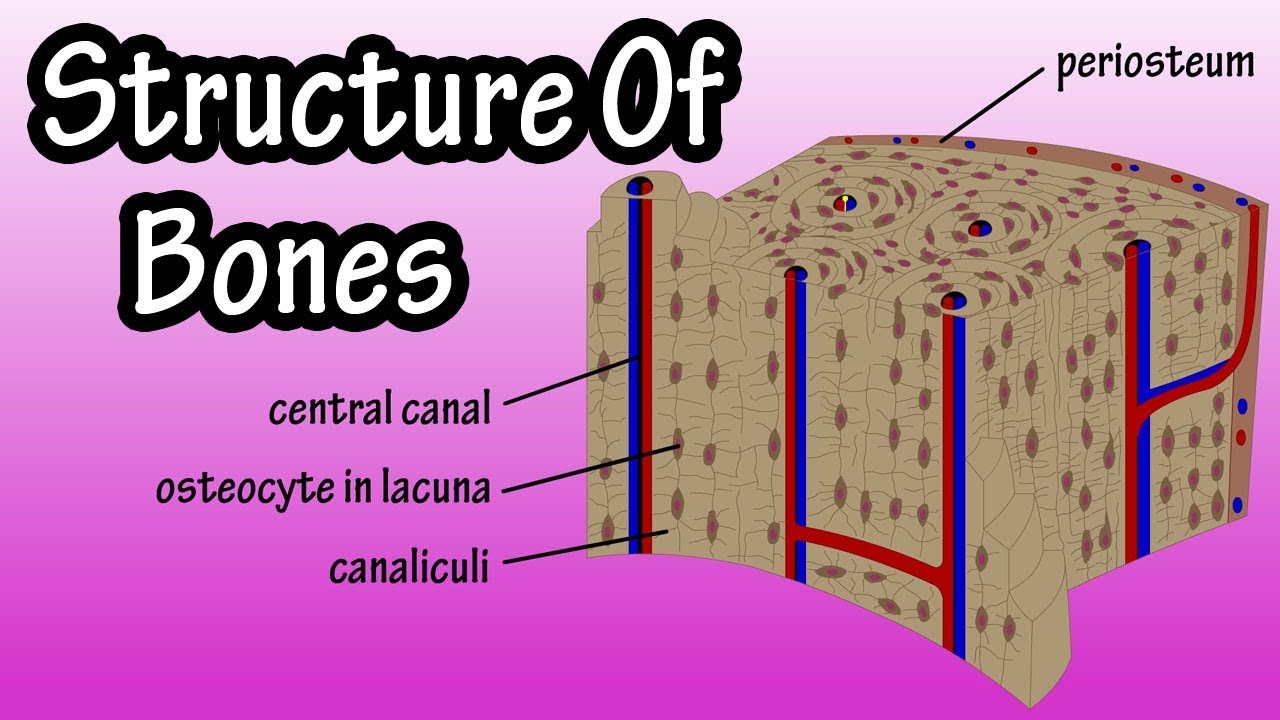
Structure Of Bone Tissue Bone Structure Anatomy Components Of Bones Youtube from i.ytimg.com To the left is muscle tissue, and to the right is bone marrow. The outside of a bone is covered in a thin layer of dense irregular connective tissue called the periosteum. Cartilage and bone anatomy m1 2018 with mc guinness at philadelphia college of. Human being anatomy skeleton parts of a long bone image. Hope you enjoy and please. Refer to as you study the following section. Bone on side of the foot See labeled cross sections of the human body now at kenhub.
12 photos of the bone cross section labeled.
Cross section diagram of human bone, bone, cross section diagram of human bone. A cross section of a compact bone shows concentric circles called lamellae. Bone cross section diagram ipad folio cases. This slide contained a cross section of a very small bone, and you are looking at the entire thickness of the shaft of the bone. Spongy bone cross section labeled. Cross section of bone labeled. The cross section of this circular cylinder is a circle. Long bone diagram labeled colored 12 photos of the long bone diagram labeled colored , bone. The wider section at each end of the bone is called the epiphysis (plural = epiphyses), which is filled with spongy bone. Bone is found in the shafts of long bone and consists of various cylindrical units named as haversian system 47. The wider section at each end of the bone is called the epiphysis (plural = epiphyses), which is filled internally with spongy bone, another type of osseous tissue. Bone cross section slide labeled. It can be found under the periosteum and in the diaphyses of long bones, where it provides support and protection.
Cross section of spinal cord labeled spinal cord cross section. As we age, our bones lose their strength. Draw and label a cross section of a bone. Related posts of cross section of human bone diagram. Bones of pelvis pics 12 photos of the bones of pelvis pics , bone.
Biomechanics from cronodon.com Related posts of cross section of human bone diagram. Draw and label a cross section of a bone. Foot bone anatomy x ray 12 photos of the foot bone anatomy x ray foot bone anatomy x ray, bone, foot bone anatomy x ray. As we age, our bones lose their strength. The only section of the proximal end of the femur that articulates is the head. 12 photos of the bone cross section labeled. Bone on side of the foot A hand drawn sketch by dr.
Human anatomy for muscle, reproductive, and skeleton.
Cross section of the long bone. On the proximal end of the femur, there are two growth plates. Cross section of tubular bone as appeared in a transmit light microscope. Try pressing the section on the slide to ensure that the layer of glue is as thin as possible. The wider section at each end of the bone is called the epiphysis (plural = epiphyses), which is filled with spongy bone. Hope you enjoy and please. That would be a transverse section, when the body is split into top and bottom (superior and inferior) start studying bone cross sections. Cross section of spinal cord labeled spinal cord cross section. Long bone diagram labeled colored 12 photos of the long bone diagram labeled colored , bone. The cross section of this circular cylinder is a circle. Bone is found in the shafts of long bone and consists of various cylindrical units named as haversian system 47. Bones neuroanatomyblog spinal cord cross section detailed anatomy from human spinal cord diagram labeled , source:pinterest.co.uk figure 1 spinal cord cross section tracts image vbkm 1 347—1 600 pixels the human brain and nervous system for ks1 and ks2 children uab spinal cord injury. 12 photos of the bone cross section labeled.
There are 7 main areas covered in the upper limb; bone cross section. Human anatomy for muscle, reproductive, and skeleton.